———— Alliance ————
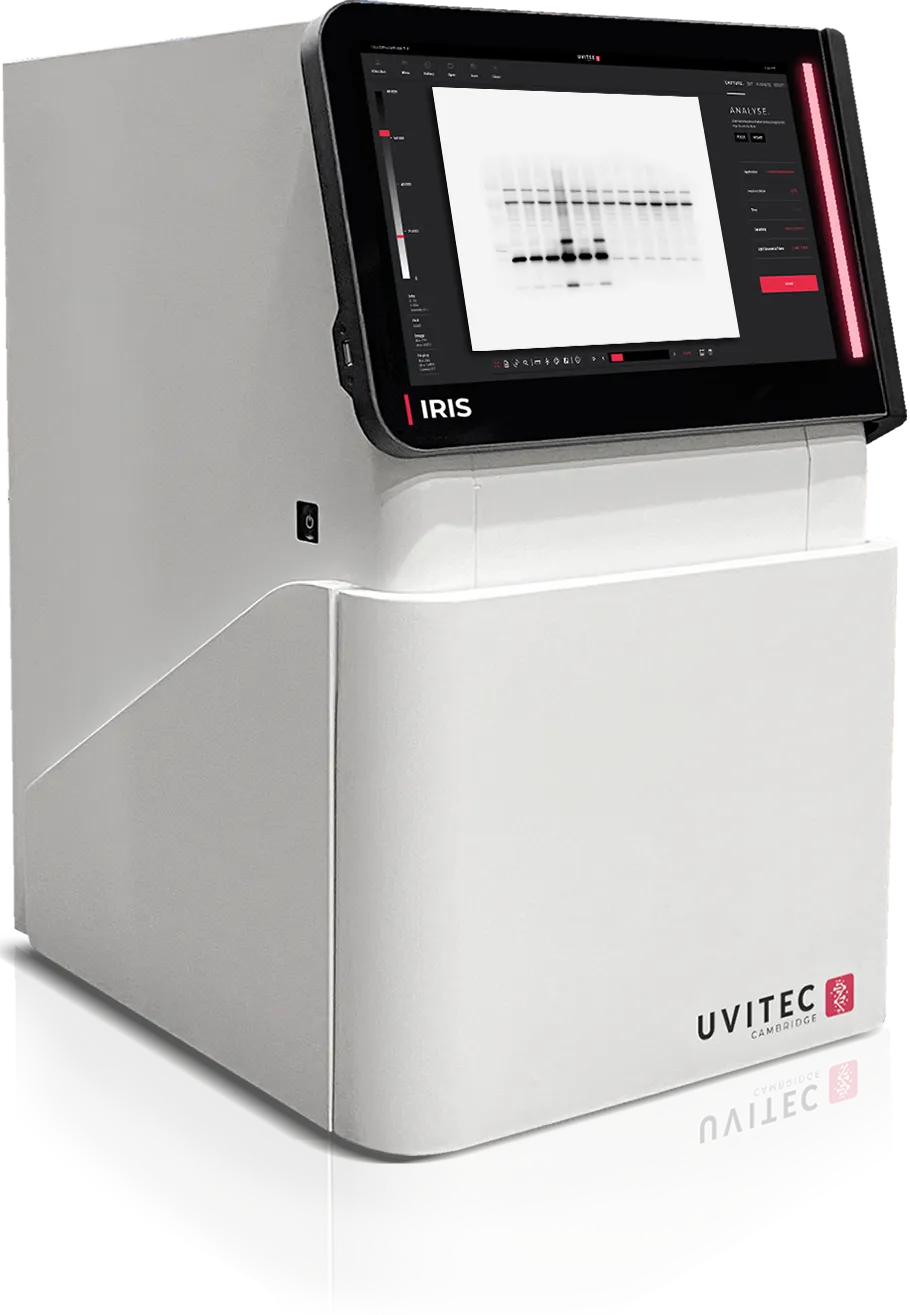
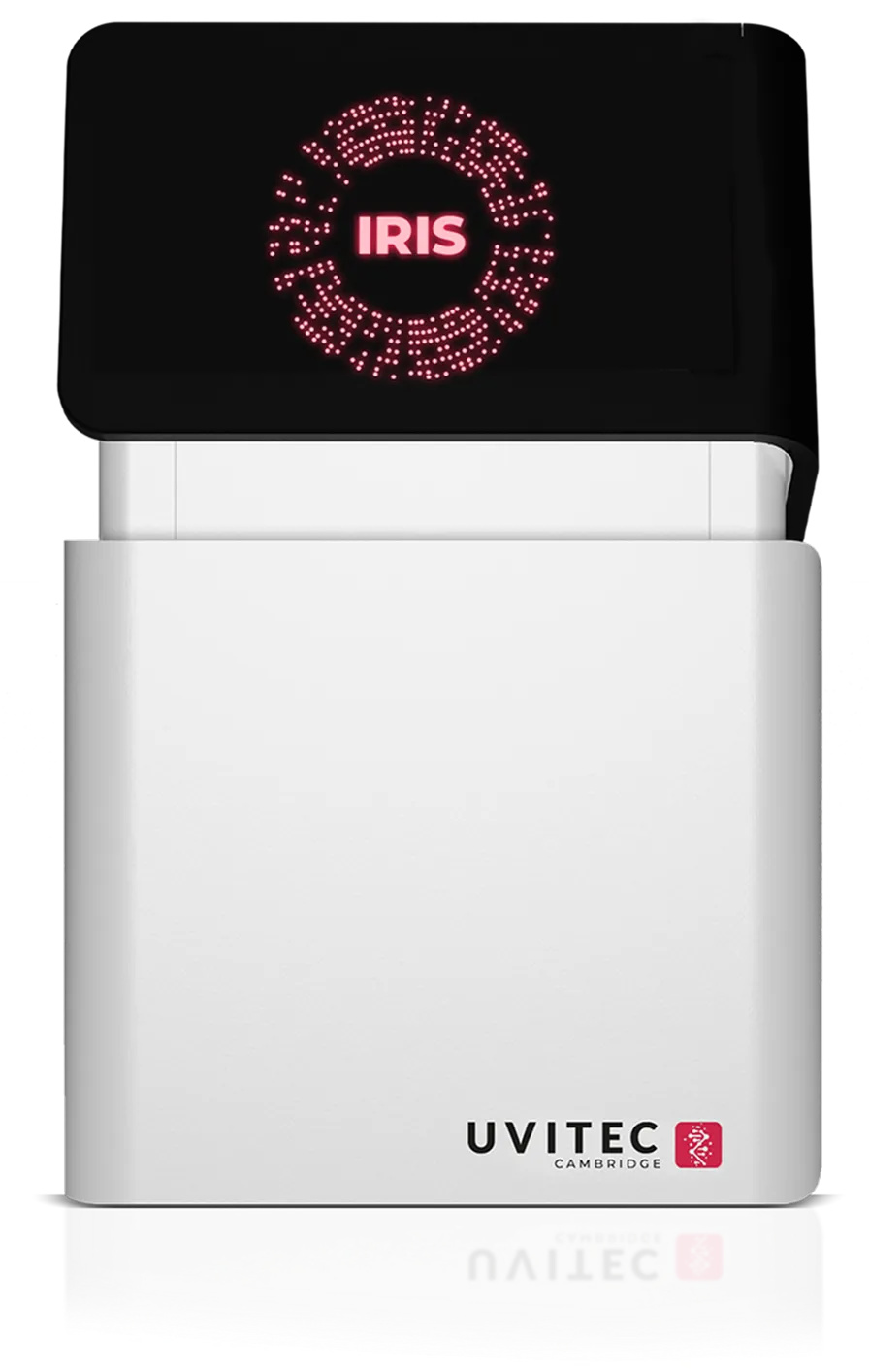
IRIS
Your Next Generation
Blot Imaging System
Alliance IRIS By UVITEC
Excellence in Every Detail
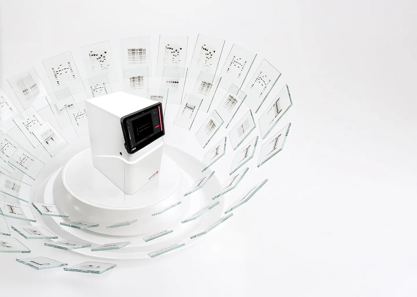
Alliance IRIS is the latest generation top-end imaging system on the market for chemiluminescent and fluorescencent Western blots. It combines outstanding optical sensitivity with advanced imaging technologies to help researchers reveal even the most challenging signals with confidence.
Developed from years of laboratory experience, every detail of the system reflects a practical understanding of scientific workflows, from image acquisition to daily usability.
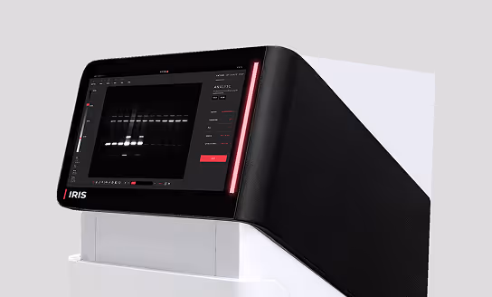
IRIS brings together elegant design, robust engineering and intuitive operation to fit naturally into modern research environments. The system is available in both touchscreen and PC-based versions for more flexibility.
LEARN MORE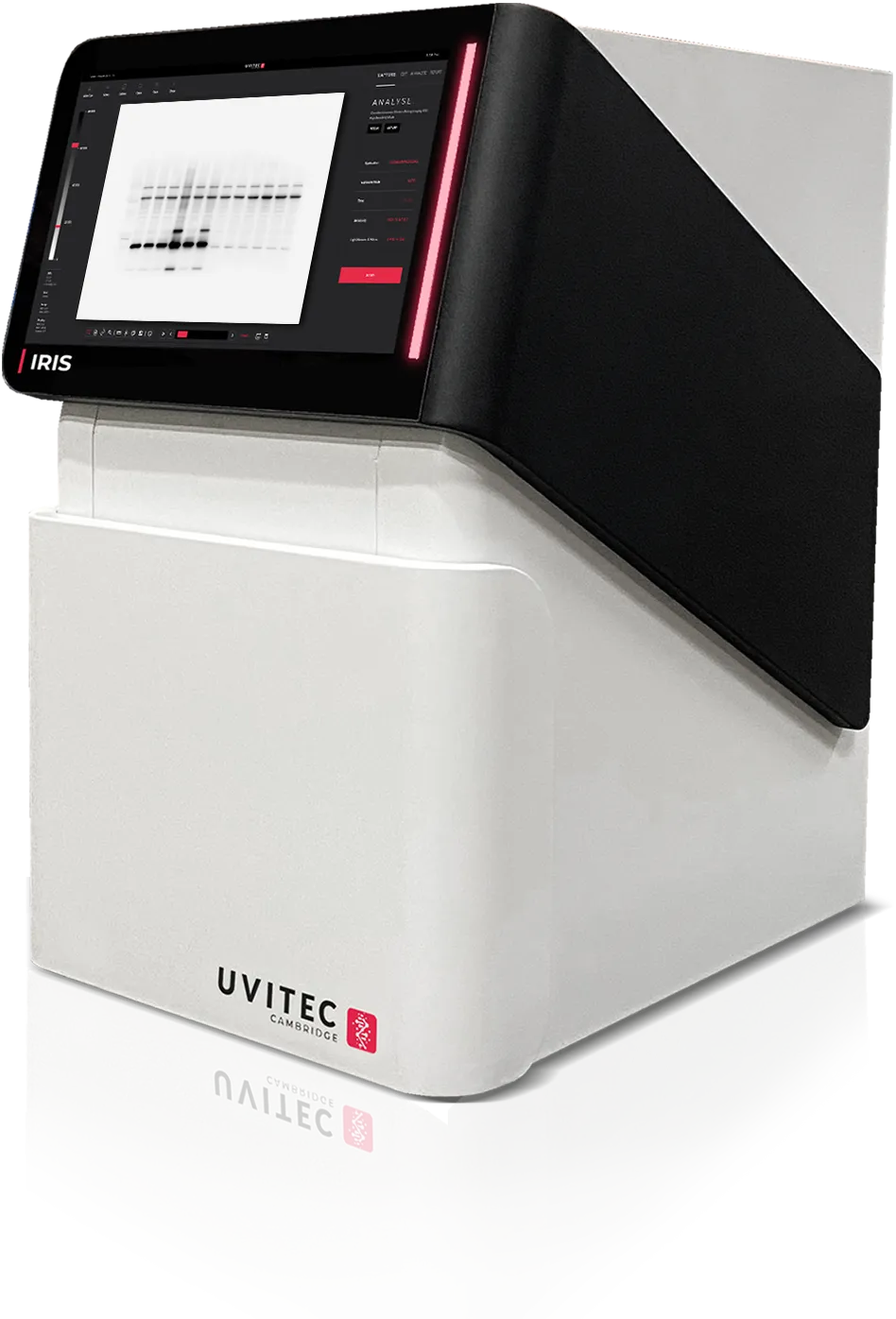
INNOVATIVE
One System,
Endless Possibilities
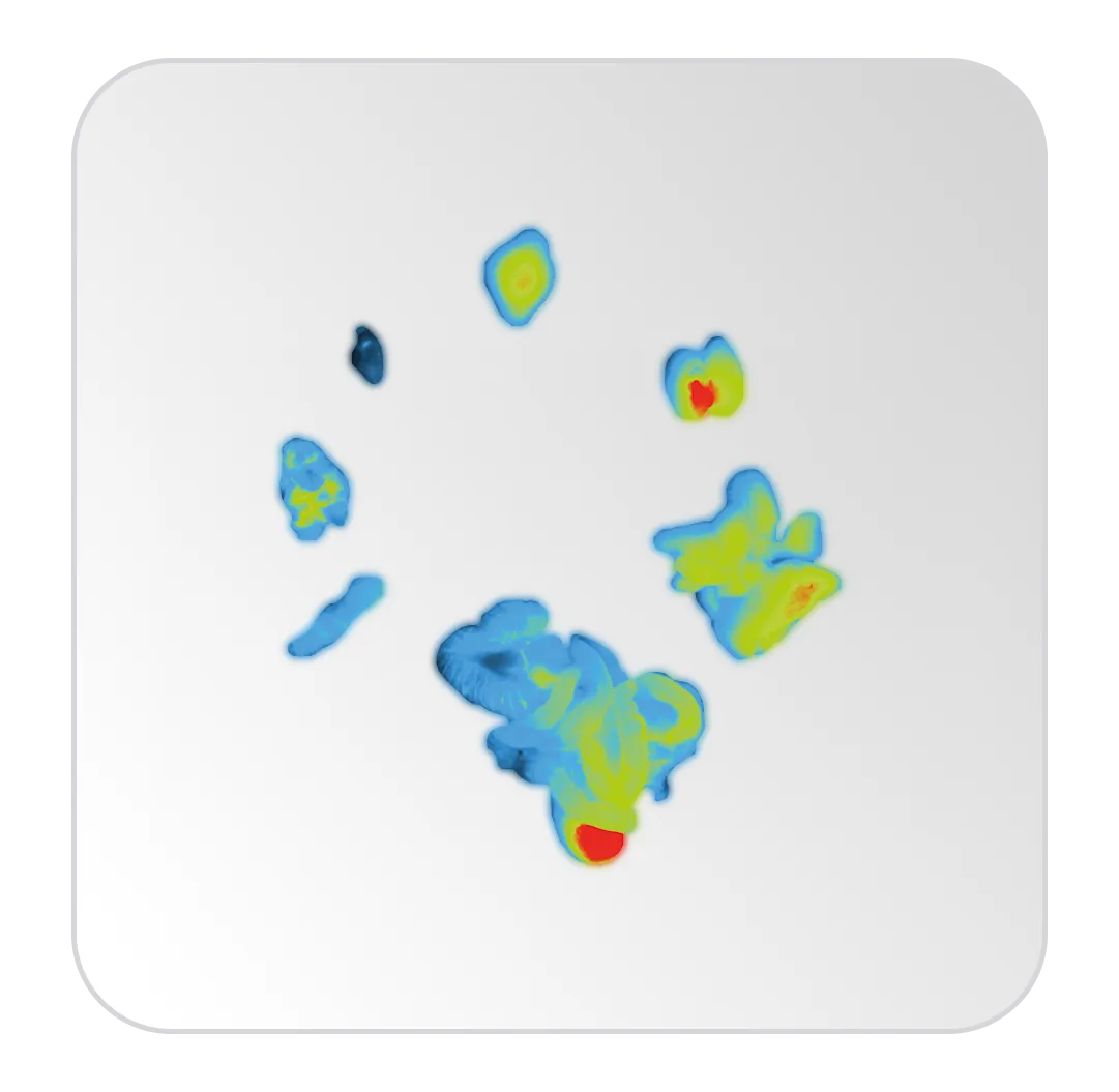
IRIS adapts to a wide range of molecular biology applications. Its modular architecture supports chemiluminescence, epi-fluorescence and UV techniques, allowing laboratories to evolve their workflows easily. To support safer and more sustainable imaging practices, IRIS also introduces a new generation of UV-LED Tables ensuring long-lasting performance and enhanced compatibility with today’s most widely used fluorescent and safe dyes.
01
Western blots
02
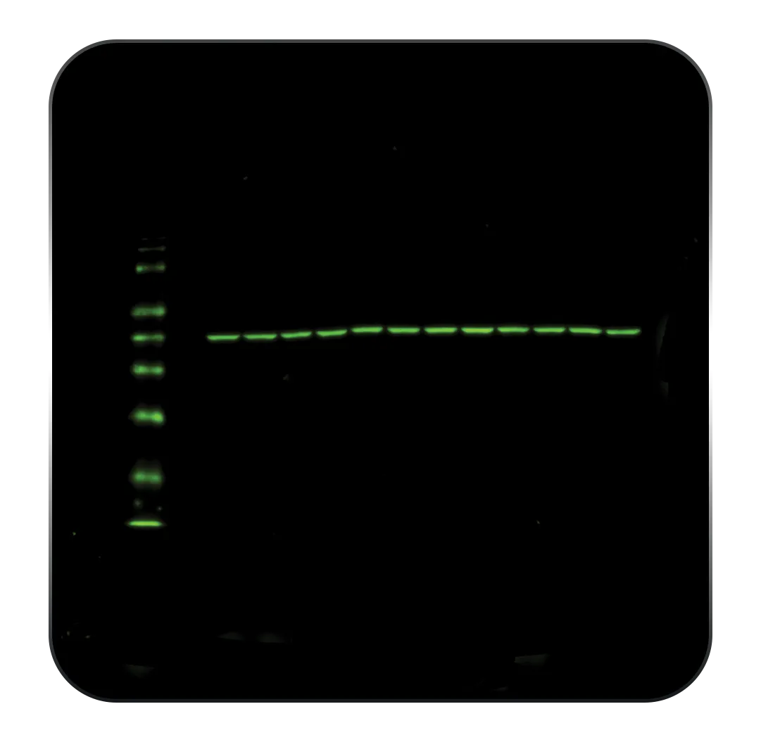
DNA / RNA gels
03
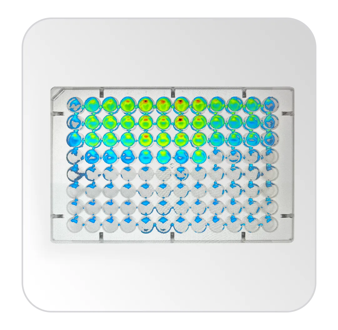
In-cell Western blots
04
Luciferase experiments
Want to check if your dyes are compatible?
LEARN MOREreliable
Uniform Illumination with
Chromascan Technology
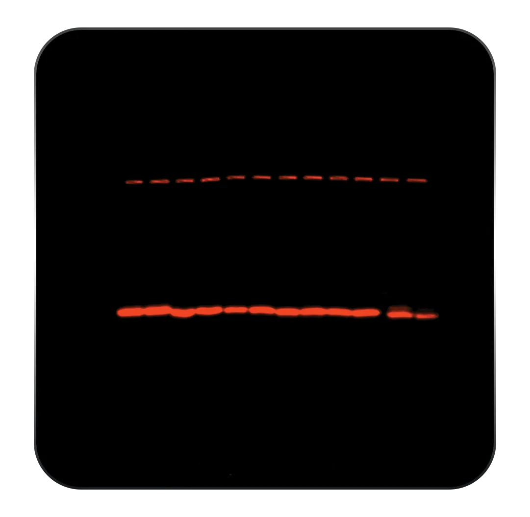
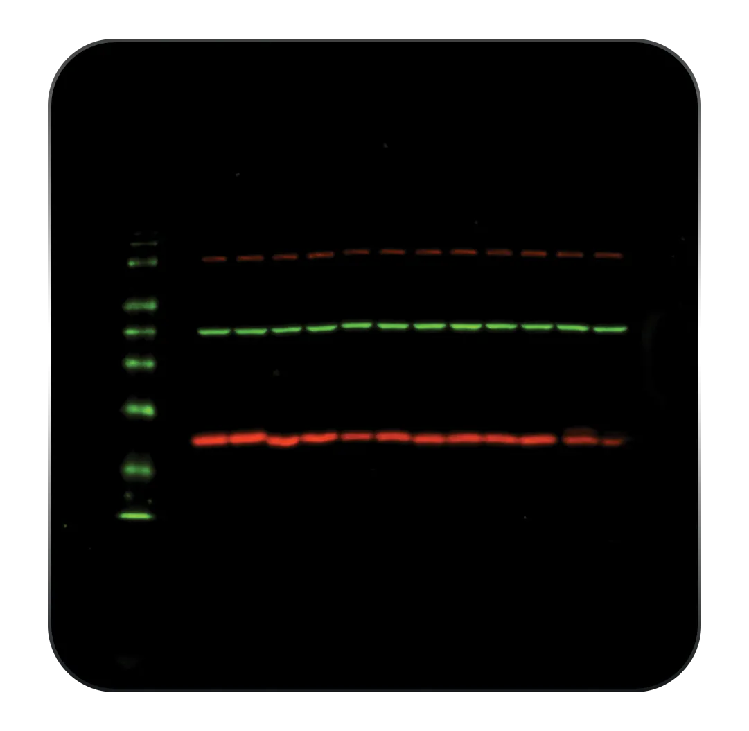
Reliable imaging begins with reliable light. IRIS integrates our advanced Chromascan illumination module, designed to deliver highly homogeneous excitation across the entire sample area. This technology combines strong excitation efficiency with exceptional multiplexing flexibility, while minimizing crosstalk between channels. IRIS supports up to 12 interchangeable excitation and emission module packs to accommodate the various numbers of epifluorescent dyes that may be used in a laboratory.
individual
ex. 680 - em. 750
+
individual
ex. 780 - em. 850
=
multiplexed
Intelligent
Effortless
Data Analysis
IRIS introduces DeepEye, an unprecedented technology in the world of molecular imaging. As soon as your Western blot is captured, our new software automatically detects lanes, bands and molecular markers - dramatically reducing the time needed for manual adjustments and allowing you to go straight to analysis. Powered by advanced image recognition and deep learning approaches, DeepEye accelerates data interpretation while preserving the integrity of raw experimental data.
sensitive
Wider
Lens Aperture
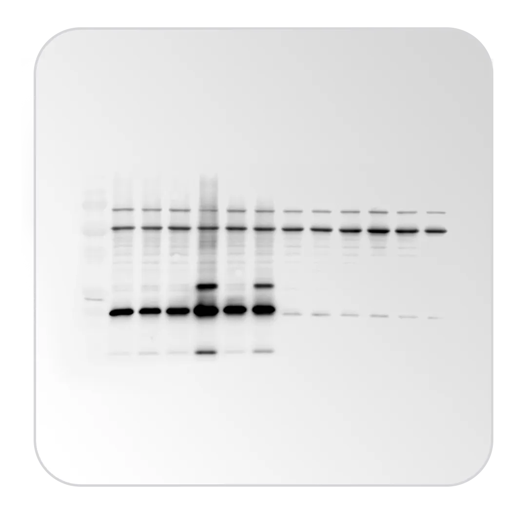
Sensitivity is at the core of our developments. IRIS’ exceptional f/0.75 aperture allows it to capture significantly more light and to detect weak protein expression and low-abundance targets with remarkable clarity. Even subtle signals remain clearly visible for reliable and confident interpretation in demanding imaging applications.
f/0.95
f/0.75
Discover the key sensitivity factors
that affect imaging performance
LEARN MOREthat affect imaging performance
CONFIGURATION
TOUCHSCREEN
Get the Cambridge Touch
Integrated standalone imaging workstation
Large adjustable 15.6” HD touchscreen
Compact footprint for optimized bench space
Robust design adapted to daily laboratory use
CONFIGURATION
PC-BASED
Built Around Your Laboratory Workflow
*PC not provided with the system
Ideal for labs with dedicated IT standards
Flexible user & data management
Convenient for shared computer setups
Familiar workflow for researchers preferring PC-based operation
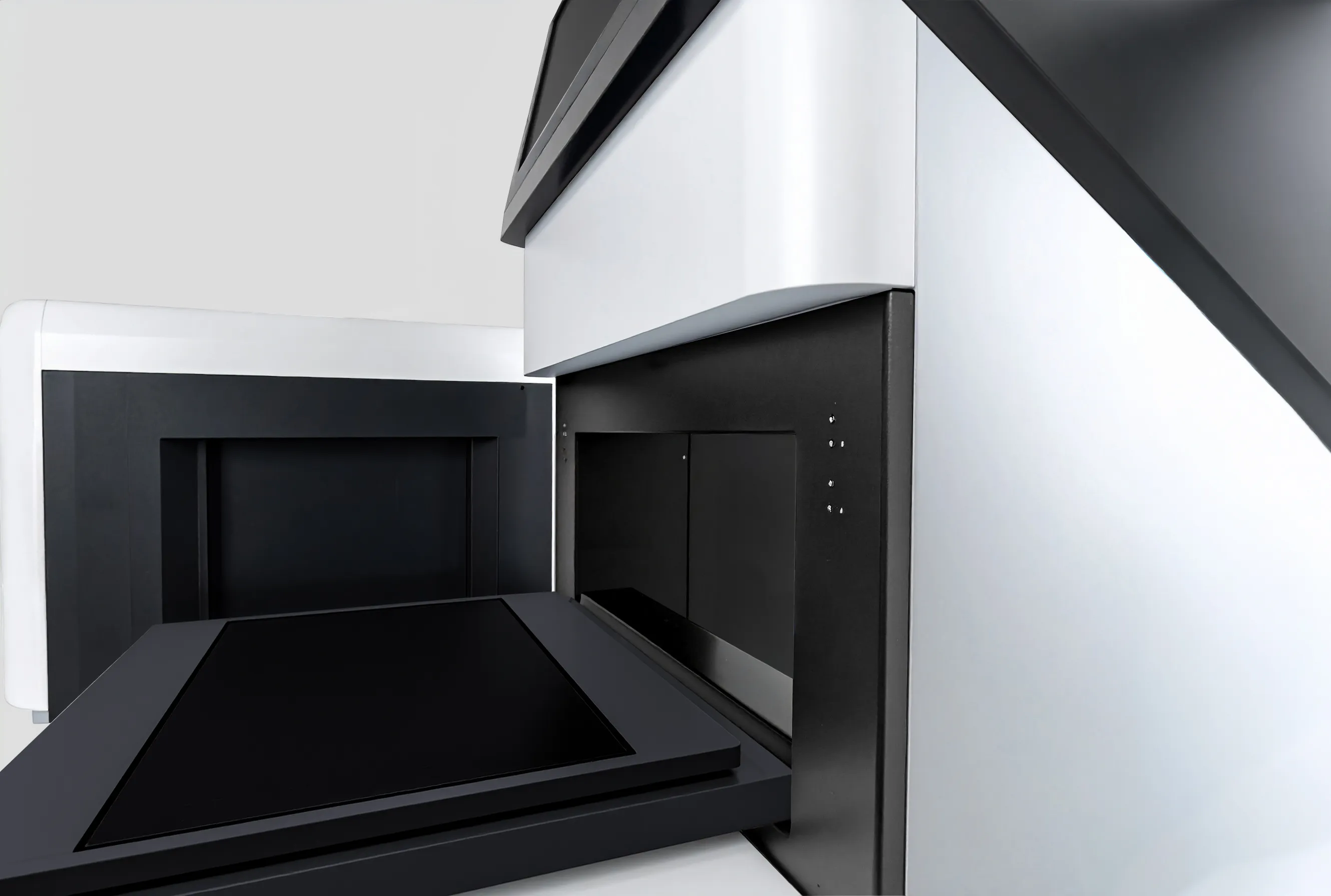
Revolutionary LED Tables
Sustainable UV-LED technology
Choice of 4 interchangeable Tables (White LED Table, Blue LED Table, 312nm UV-LED Table and Black Table for chemiluminescence and epifluorescence)
Plug n’ Play for effortless sample positioning
Uvipure for enhanced UV for EtBr and all safe stains
Innovative Chromascan concept
Homogeneous illumination across your sample
Multiplexing possibilities
No crosstalk issues
12 Chromascan modules packs available from 365nm to 780nm (including a combination of excitation light sources and emission filters)
Chromascan Modules
Application Dyes
Chromascan UV365 pack module
EX365nm-EM590nm
EX365nm-EM590nm
Qdot 565, Qdot 655, Qdot 705, TLC plates, microplate, Alexa 350, DAPI
Chromascan Light Blue pack module
EX440nm-EM500nm
EX440nm-EM500nm
DAPI, CFP, Cerulean, Alexa Fluor 405, Cascade Blue, Pacific Blue, DyLight 405, Atto 425
Chromascan Blue pack module
EX480nm-EM550nm
EX480nm-EM550nm
FITC, Alexa Fluor 488, GFP, SYTOX Green, Fluorescein, Cy2, Sypro Ruby, DyLight488
Chromascan Deep Blue pack module
EX480nm-EM600nm
EX480nm-EM600nm
YFP, eYFP, Venus, Alexa Fluor 514, FITC, mCitrine
Chromascan Green pack module
EX540nm-EM600nm
EX540nm-EM600nm
Rhodamine, Alexa Fluor 532, Alexa Fluor 555, Cy3, PE, TRITC, ProQdiamond, DyLight 549, mRuby3
Chromascan Deep Green pack module
EX540nm-EM650nm
EX540nm-EM650nm
Cy3.5, Atto 565, Rhodamine 6G
Chromascan Orange pack module
EX580nm-EM650nm
EX580nm-EM650nm
DsRed, mCherry, Cy3.5, Alexa Fluor 568, Texas Red, Atto565, Atto594, Alexa594, mStrawberry, mKate2
Chromascan Red pack module
EX640nm-EM700nm
EX640nm-EM700nm
Alexa Fluor 647, Alexa Fluor 660, Cy5, APC, Atto633, Atto 647N, DyLight 633, DyLight 650
Chromascan NIR pack module
EX680nm-EM750nm
EX680nm-EM750nm
Alexa Fluor 680, Alexa Fluor 700, Cy5.5, IRDye 680RD, Atto 680, Atto 700, APC-Cy7, DyLight 680
Chromascan LIGHT IR pack module
EX740nm-EM750nm
EX740nm-EM750nm
Alexa Fluor 750, Cy7, IRDye 800CW, VivoTag-S 750, DyLight 755
Chromascan IR pack module
EX740nm-EM800nm
EX740nm-EM800nm
Cy7, IRDye 800RS, ZW800-1, Atto 740, HiLyte Fluor750
Chromascan FAR IR pack module
EX780nm-EM850nm
EX780nm-EM850nm
Alexa Fluor 790, Cy7.5, IRDye 800CW, VivoTag-S 800, DyLight 800
Extreme sensitivity
Massive resolution of 9.2 megapixels for HD pictures
f/0.75 custom lens and unrivalled camera sensitivity
Absolute cooling of -30°C via 3 stages Peltier
Outstanding weak/strong detection ratio with OD 4.8 dynamic range
Autofocus motorized optics
Femtogram-level detection
High-end design
Stainless steel and epoxy paint for long-term robustness
Standalone system with integrated PC
Widest 15.6 inches touch screen
Dimensions: 670 mm (height) x 435 mm (width) x 540 mm (depth)
Weight: 70 Kg